
Clinical history:
25 year old patient with a positive pregnancy test and last menstrual period 8 weeks prior.
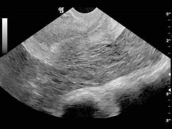
This sagittal transvaginal ultrasound image shows:
- An abnormally thickened endometrial stripe
- A normal intrauterine pregnancy
- Uterine leiomyomas
- Absence of a gestational sac
What finding in this image is of greatest concern in a patient with the given history?
The yolk sac is normally visible at gestational age of:
- 4 weeks
- 5.5 weeks
- 8 weeks
- 10.5 weeks
What value of quantitative beta HCG is used as a guideline for when a normal intrauterine pregnancy should be apparent by transvaginal ultrasound?
- 1,000
- 2,000
- 10,000
- 20,000
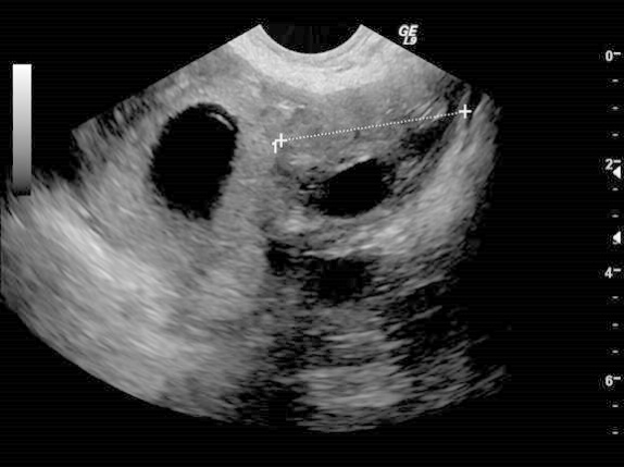
Identify the two hypoechoic areas above.
What finding in the above image is indicative of a tubal ectopic pregnancy?
- The double decidual sign
- The presence of a pseudogestational sac
- The tubal ring sign
- The ring of fire sign
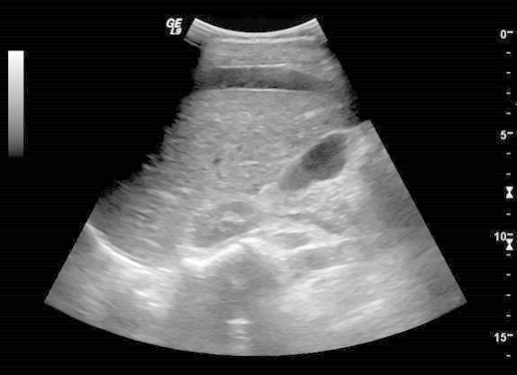
An additional transabdominal image taken at the patient’s right upper quadrant shows:
- Liver infarction due to HELLP syndrome
- Hepatic steatosis
- Hemoperitoneum
- Simple ascites
- Pneumoperitoneum
- This image shows the absence of a gestational sac.
- Posterior to the uterus is a collection of hyperechoic free fluid, indicative of hemorrhage.
- The yolk sac is typically visible by 5.5 weeks.
- With a quantitative beta HCG of 2000 an intrauterine pregnancy, when present, is usually seen on transvaginal ultrasound.
- The structure on the right of the image is a normal ovary. The structure on the left is a tubal ectopic gestational sac.
- The tubal ring sign in this image is indicative of a tubal ectopic pregnancy.
- This image shows perihepatic fluid from a large amount of hemorrhage secondary to the tubal pregnancy.
- This additional transverse image demonstrates a gestational sac separate from the uterus:
Discussion:
A conservative estimate of the rate of ectopic implantation of an embryo according to the CDC is approximately 2% of all pregnancies. As ectopic pregnancy is the leading cause of pregnancy related fatality during the first trimester, it is important to be able to diagnose the condition in a timely fashion. Furthermore, accuracy in diagnosis is essential to avoid the loss of a desired normal intrauterine pregnancy due to misdiagnosis as an ectopic pregnancy. Patients with an ectopic pregnancy often present with complaints of abdominal pain, vaginal bleeding, and amenorrhea. Though these symptoms are somewhat nonspecific, initial evaluation should include studies aimed at ruling out an ectopic pregnancy. Several risk factors have been identified, such as previous ectopic pregnancy, history of pelvic inflammatory disease, use of an intrauterine device, and a history of tubal or other gynecologic surgery. However ectopic implantation often occurs in the absence of any risk factors. A quantitative beta HCG level should be obtained. Given the variability in patients’ ability to report an accurate date of last menstrual period, the reported level is necessary to interpret the findings or lack of findings upon scanning. After establishing the stability of the patient, transabdominal and transvaginal sonography may be performed. Initial transabdominal scanning is useful for visualization of the superior portion of the uterus and for evaluation of adnexa which are futher superior in the abdomen. More importantly, the degree of hemoperitoneum, when present, can be assessed. If a significant amount of hemoperitoneum is found, depending on the stability of the patient, the remainder of the exam should be carried out as quickly as possible or alternatively discontinued in order to proceed with urgent surgical intervention. Using transvaginal sonography, in normal intrauterine pregnancies, the intradecidual sign can be seen at 4.5 weeks after the last menstrual period. The double decidual sign should be apparent by 5 weeks gestation. A secondary yolk sac may be seen at 5.5 weeks. By 5-6 weeks cardiac activity should be detectable. A beta-HCG level of 2000 mIU/mL is used as the discriminatory guideline at which an intrauterine pregnancy, when present, will be apparent by transvaginal ultrasound. Note that the level can vary by institution depending on the assay used. Even if the level is found to be below the cutoff value, sonography should still be performed as it is often possible to define a normal intrauterine pregnancy. However, if no intrauterine pregnancy is identified by sonography in a patient with a level greater than 2000, there should be a high suspicion of ectopic pregnancy. Additionally, even if a normal intrauterine pregnancy is established, a second ectopic pregnancy might be considered particularly in high risk patients. Heterotopic implantation can occur in up to 1 of 4000 normal pregnancies and as many as 1 of 100 pregnancies resulting from in vitro fertilization techniques. Care should be taken not to mistake a pseudogestational sac for a true intrauterine pregnancy. A pseudosac is surrounded by only a single layer of hyperechoic tissue. This is in contrast with the finding of two hyperechoic rings surrounding the gestational sac in a normal intrauterine pregnancy – a feature known as the double decidual sign. The psuedogestational sac may contain heterogeneous material but can be further distinguished from a true gestational sac by its central location within the endometrial canal. The most specific finding of ectopic pregnancy is, not surprisingly, the visualization of an extrauterine gestational sac, or failing that, an adnexal mass. Most (95%) ectopic pregnancies occur in the ampulla or isthmus of the fallopian tube. Other types of abnormal implantation include interstitial, and more rarely intramural, abdominal, cervical, ovarian, and along cesarean section scars. A tubal pregnancy may appear as a hyperechoic tubal ring containing an embryo, a yolk sac, a tubal ring without any identifiable contents, or a complex adnexal mass which is not within the ovary. If a complex mass is identified within the ovary, it is very unlikely to be an ectopic pregnancy, as implantation in this location is rare, and is most likely a corups luteum cyst. Using gentle pressure with the transducer can help to differentiate a mass which moves with the ovary versus one that moves separately from the ovary. Unfortunately, the use of color Doppler to look for the “ring of fire” sign, which describes a rim of increased blood flow around an adnexal mass, cannot definitively differentiate a tubal pregnancy from a corpus luteum cyst. If a case arises in which no intrauterine or ectopic pregnancy is found in a stable patient with a positive beta-HCG, the patient may be managed expectantly with follow up quantitative beta-HCG levels and ultrasound within 2-3 days. Treatment options for an ectopic pregnancy depend heavily on the clinical scenario. Surgical intervention, whether by laparotomy or laparoscopy, is necessary in the management of unstable patients. Those who are stable at the time of presentation and who are expected to be reliable regarding follow up may be treated medically with methotrexate injection. Some patients will be treated via sonographically guided percutaneous treatment, if the location or the presence of an additional intrauterine pregnancy warrants directed intervention. Occasionally stable patients will also be managed expectantly and followed with serial beta-HCG values and sonography. Deirdre Gallagher, M.D. University of Pittsburgh Medical Center Department of RadiologyReferences:
- Jurkovic D. Mavrelos D. Catch me if you scan: ultrasound diagnosis of ectopic pregnancy. Ultrasound in Obstetrics & Gynecology. 30(1):1-7, 2007 Jul.
- Barnhart KT. Simhan H. Kamelle SA. Diagnostic accuracy of ultrasound above and below the beta-hCG discriminatory zone. Obstetrics & Gynecology. 94(4):583-7, 1999 Oct.
- Lin EP. Bhatt S. Dogra VS. Diagnostic clues to ectopic pregnancy. Radiographics. 28(6):1661-71, 2008 Oct.
- Levine D. Ectopic pregnancy. Radiology. 245(2):385-97, 2007 Nov.
- Fleischer AC. Pennell RG. McKee MS. Worrell JA. Keefe B. Herbert CM. Hill GA. Cartwright PS. Kepple DM. Ectopic pregnancy: features at transvaginal sonography. Radiology. 174(2):375-8, 1990 Feb.
- Pellerito JS. Taylor KJ. Quedens-Case C. Hammers LW. Scoutt LM. Ramos IM. Meyer WR. Ectopic pregnancy: evaluation with endovaginal color flow imaging. Radiology. 183(2):407-11, 1992 May.
- Barnhart KT. Sammel MD. Rinaudo PF. Zhou L. Hummel AC. Guo W. Symptomatic patients with an early viable intrauterine pregnancy: HCG curves redefined. Obstetrics & Gynecology. 104(1):50-5, 2004 Jul.
- Seeber BE. Barnhart KT. Suspected ectopic pregnancy. Obstetrics & Gynecology. 107(2 Pt 1):399-413, 2006 Feb.
- Hammoud AO. Hammoud I. Bujold E. Gonik B. Diamond MP. Johnson SC. The role of sonographic endometrial patterns and endometrial thickness in the differential diagnosis of ectopic pregnancy. American Journal of Obstetrics & Gynecology. 192(5):1370-5, 2005 May.
- Doubilet PM. Benson CB. Frates MC. Ginsburg E. Sonographically guided minimally invasive treatment of unusual ectopic pregnancies. Journal of Ultrasound in Medicine. 23(3):359-70, 2004 Mar.