
Clinical history:
54 year old woman with right upper quadrant pain
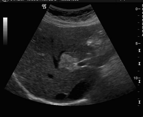
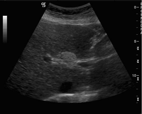
The diagnosis of this hyperechoic hepatic lesion is?
- Hepatocellular carcinoma
- Focal fatty infiltration
- Hepatic adenoma
- Metastatic disease
Is this a typical location for this lesion?
This lesion can be associated with the following:
- Alcohol abuse
- Diabetes mellitus
- Pregnancy
- Steroid therapy
- Chemotherapy
- Alcohol abuse, diabetes, and pregnancy
- None of the above
- All of the above
What tests can be used to confirm the diagnosis?
Discussion:
Fatty infiltration of the liver represents abnormal triglyceride uptake within hepatocytes and may be either diffuse or focal. Focal fatty infiltration is a relatively common entity and is well recognized both radiologically and histopathologically. Whereas focal infiltration of the liver can often be distinguished from space occupying lesions of the liver, when it presents in a nodular form diagnosis can be difficult. The differential diagnosis includes other hyperechoic hepatic lesions including hepatocellular carcinoma, metastasis, and cavernous hemangiomas. Focal nodular hyperplasia and hepatic adenoma are less likely possibilities.
Focal fatty infiltration is most common within the anterior aspect of the medial segment of the left lobe of the liver (segment IV), immediately adjacent to the falciform ligament. Another common location is anterior to the portal vein bifurcation. In addition to these customary locations, there are other typical findings which help distinguish focal fatty infiltration from the previously mentioned hyperechoic hepatic lesions. The boundaries between the focal fatty hepatocytes and normal liver parenchyma may have interdigitating and geographic margins (although a nodular configuration is not uncommon). Fatty infiltration tends not to cause contour abnormalities on adjacent hepatic and portal veins, even to the extent of allowing branches to pass through the fatty lesion undisplaced. In addition, a rapid change with time is characteristic, with near complete resolution after nutritional improvement in as little as a few days.
Fatty infiltration of the liver is a pervasive finding seen in a variety of patient populations. Focal infiltration is commonly associated with alcohol abuse and morbid obesity, yet may also be seen in conjunction with diabetes mellitus, pregnancy, steroid therapy, chemotherapy, malnutrition and hyperalimentation. Whereas the exact etiology of this focal fatty deposition remains a point of contention, it is assumed to be related to regional differences or disturbances in hepatic portal blood flow. Other theories as to the focality of this fatty deposition involve regional tissue hypoxia and focal accumulation of toxins.
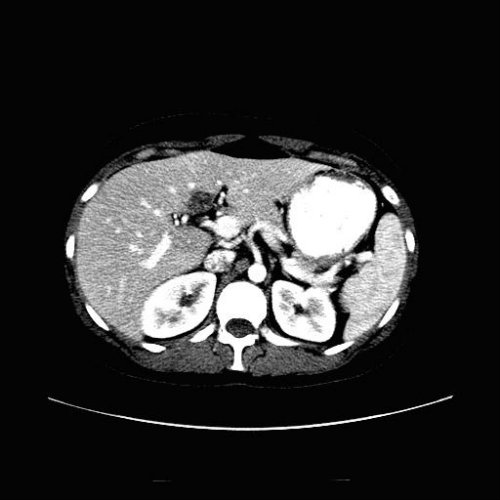
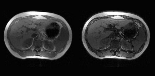
If a hyperechoic lesion is seen in these classic locations, in a patient without a history of a known primary malignancy, the diagnosis is most likely focal fatty infiltration and no further evaluation is warranted. However, in a patient with a history of malignancy, or in the presence of a hyperechoic lesion with an atypical shape or location, a subsequent study may help to confirm the diagnosis. MRI with in- and out-of-phase sequences and non- contrast computed tomography are commonly used in equivocal cases. When the diagnosis remains uncertain, invasive procedures such as percutaneous biopsy are then performed.
Cory Nordman, M.D.
Department of Radiology, University of Pittsburgh Medical Center
References:
- Yoshikawa, Matsui, Takashima, Sugiura, Katayama, Nishida, Tsuji; Focal Fatty Change of the Liver Adjacent to the Falciform Ligament: CT and Sonographic Findings in Five Surgically Confirmed Cases; AJR 149:491-494, September 1987.
- S Quinn, B Gosink; Characteristic Sonographic Signs of Hepatic Fatty Infiltration; AJR 145: 753-755, October 1985.
- M Baker, J Wenker, E Cockerill, J Ellis; Focal fatty infiltration of the live: Diagnostic imaging; Radiographics Vol 5, Number 6, November 1985.
- J Kemper, G Jung, W Poll, C Jonkmanns, R Luthen, U Moedder; CT and MRI findings of multifocal hepatic steatosis mimicking malignancy; Abdominal Imaging 27:708-710, 2002.
- H Tamai, et al; Mutitfocal Nodular Fatty Infiltration of the Liver Mimicking Metastatic Liver Tumors; J Ultrasound Medicine 25:403-406, 2006.
- Middleton W, Alfred K, Hertzberg B. The Requisites: Ultrasound, second edition; 2004. Mosby.v