
Clinical history:
59 year old male with “clicking” of right 4th digit with extension
The most likely diagnosis is:
- Ganglion cyst
- Trigger finger
- TenosynovitisGout
The treatment for this condition includes:
- Steroid injections
- Splinting
- Surgery
- All of the above
Answers: b, d
Sonographic findings:
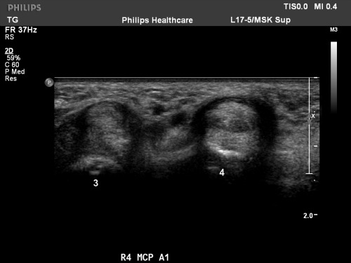
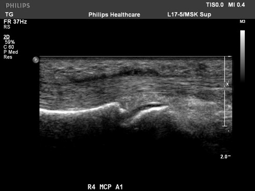
A1 pulley is continuous with the tendon sheath, proximal to the MCP joint. The 3rd digit in figure 1 demonstrates normal anatomy as imaged with a high-frequency (17MHz) probe. The A1 pulley is a thin, hypoechoic connective tissue band (black arrows) that overlies the flexor tendons (FT) and volar plate (‘*’) at the level of the MCP joint. The abnormal 4th digit demonstrates focal hypoechoic thickening of the A1 pulley (white arrows). In figure 2, there is also focal decreased echogenicity and thickening of the flexor tendons (white arrowheads), likely due to underlying tendinosis.
Other sonographic findings of trigger finger include swelling of the flexor tendons, synovial sheath effusions and hypervascularization of the A1 pulley on power Doppler. On longitudinal scanning with flexion, the tendon catches during movement and gliding movement is not smooth
Discussion:
Normal finger flexion is orchestrated in part by the flexor pulley system–focal thickened areas of the fibrous flexor tendon sheath. The pulley system comprises the annular and cruciform systems. The annular system is formed by thick, transverse fibers, located over the metacarpophalangeal and proximal and distal interphalangeal joints and the midshaft of the proximal and middle phalanx. The cruciform system is formed by thin, criss-crossing fibers that interconnect the annular system. The pulleys affix the flexor tendons to the phlangeal cortex and provide the points at which the force of the tendons is exerted during flexion. Injury to the pulley system causes volar bowstringing, or anterior displacement of the flexor tendons, and reduces digital performance. Tendinopathy along with thickening of the A1 pulley specifically leads to symptoms of trigger finger. Symptoms of trigger finger can range from tightness or pain in the hand to locking and triggering of the involved digit. Pathophysiologically, symptoms are caused by mismatch between the relative size of the flexor tendon and its sheath by various conditions involving the tendon or its sheath or both.Though incidence is increased in patients with diabetes, rheumoatoid arthritis, De Quervain’s tneosynovitis, osteoarthritis and hypothyroidism, most trigger fingers have no recognizable cause. Trauma to the tendon is a rare cause. The diagnosis is made clinically, and first-line treatment is ultrasound-guided corticosteroid injection and splinting of the finger. Surgical intervention with secitoning of the A1 pulley may be indicated in some instances.
References:
- Bodor M, Flossman T. Ultrasound-guided first annular pulley injection for trigger finger. J Ultrasound Med; 28: 737-743.
- Boutry N, Titecat M, Demondion X, Glaude E, Fontaine C, Cotten A. High-frquency ultrasonographic examination of the finger pulley system. J Ultrasound Med 2005; 23: 1333-1339.
- Guerini H, Pessis E, Theumann N, Le Quintrec JS, Campagna R, Chevrot A, Feydy A, Drape JL. Sonographic appearnace of trigger fingers. J Ultrasound Med 2008; 27: 1407-1413.
- Serafini G, Derchi LE, Piergiorigio Q, Martinoli C, Orio O, Cavallo, Gandolfo N. High resolution sonography of the flexor tendons in trigger fingers. J Ultrasound Med 16; 213-219.